Orbital Floor or Blowout Fractures
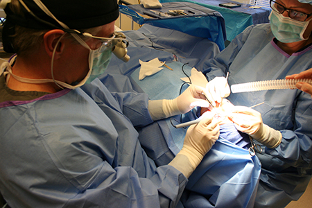
Some of the bones surrounding the eye (in the eye socket or orbit) are extremely thin and delicate. These bones are therefore easily fractured when the orbital area is traumatized. This kind of trauma to the orbital area is common in motor vehicle accidents as well as in assaults. A sudden increase in the pressure in the eye socket or orbit may in itself cause these delicate bones to fracture into the surrounding sinus cavities, either below or on the nasal side of the eyes. Buckling of the stronger bones around the eye such as the maxilla or cheek bone may also contribute to fracture of the delicate orbital bones. When an orbital floor or blowout fracture occurs, soft tissue content of the orbit can become trapped in the fracture site on the floor of the eye socket resulting in restriction of the movement of the eye. This can result in double vision (called diplopia in medical language) especially when looking up. The movement of soft tissue (primarily fat) content of the orbit or eye socket through the fractured bone to the surrounding sinuses can also cause the eyeball to sink back (enophthalmos) or sink down (hypoglobus) in the eye socket. This change in eyeball position may become apparent very slowly as the swelling related to the injury resolves. This shifting of the eye position following trauma can take up to one year. The larger the fracture, the more likely it is that the eye will move out of position. Surgical repair of orbital floor “blowout fractures” often involves the use of implant material to cover the fracture site after the orbital tissues (primarily fat but also at times muscle) are returned to their natural position in the orbit. Dr. Popham most often repairs this kind of fracture through an incision on the inside of the eyelid, thus avoiding any sutures or scarring on the skin.
Orbital Tumors
Both benign and malignant (or cancerous) tumors may occur in the eye socket or orbit. Such tumors may begin within the orbit while others begin in the sinuses or brain and extend into the eye socket. Occasionally, an orbital tumor may come from a cancer elsewhere in the body in a process called metastasis. The most common tumors of the eye socket in adults are composed of blood vessel elements and are benign. The most common tumors in the eye socket in children are also benign and may be composed of skin related structures or of blood vessels. Orbital tumors may cause restriction of the movement of the eye and thereby cause double vision or diplopia. Orbital tumors may also push the eyeball forward or out of position. Other symptoms may include pain, loss of vision, and swelling around the eye. If an orbital tumor is suspected, Dr. Popham may order a CAT scan or an imaging study called an MRI. Many orbital tumors require biopsy for diagnosis.
Thyroid Eye Disease or Grave’s Disease
This disease is generally thought to be an autoimmune disease, or a disease in which the patient’s own immune system attacks an otherwise normal body structure. This disease has been referred to as Grave’s disease, thyroid eye disease (TED), thyroid related orbitopathy (TRO), Grove’s orbitopathy (GO), etc. Thyroid eye disease affects females more than males at a ratio of about 8:1. Many patients with Grave’s disease have family members who have experienced the same. Grave’s disease classically refers to a disease state in which the patient’s immune system attacks receptors on the thyroid gland, causing the thyroid gland to over produce thyroid hormones (hyperthyroidism). This typically causes the patient to experience symptoms such as a rapid heartbeat, weight loss, hair loss, overheating, tremors, and irritability. Physical findings may include goiter, or an enlarged thyroid gland in the neck area. Smoking cigarettes aggravates the eyelid and orbital effects of Grave’s disease. The eye and orbital effects of thyroid eye disease include proptosis (a forward displacement or bulging of the eye or eyes), double vision, and eyelid retraction (the upper eyelids may pull up and the lower eyelids may pull down). In a worst case scenario, thyroid eye disease may enlarge the eye movement muscles which may squeeze the optic nerve. If this compression of the optic nerve (compressive optic neuropathy), persists or is severe, it can restrict blood flow to the nerve or the eye and cause loss of vision. The inflammatory components of Grave’s disease are often treated initially with anti-inflammatory medications or other medications. Radiation is sometimes used to control the inflammatory effects of the disease. Surgery may be needed to correct the negative effects of Grave’s disease. Surgeries are most commonly performed after the disease has become quiescent. Such surgeries may include orbital decompression (opening thin bones and removing fat in the eye socket to take pressure off the eye and optic nerve), eye muscle (strabismus) surgery (surgical repositioning of the eye movement muscles done to decrease or correct double vision or diplopia), or eyelid retraction surgery (moving the upper eyelids down or moving the lower eyelids up).

